
 Just when you think you've seen all the crystals you ever want to see, ju
 st take a look at these. These are uric acid crystals from a finger joint.
Exhibiting all of their full wave plate color compensated polarizing charaacteristics.
Now here are some pics from a knee fluid.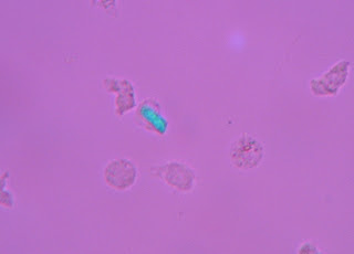
 These are calcium pyrophosphate crystals.

 These are calcium pyrophosphate crystals.
 Okay are you ready for this? Intracellular AND extracellular (in the same field!!) (This picture could be better, but really I was very busy)

And just when you think you've seen everything (or at least more than you wanted to see)...
I have one more. Earlier in the day I commented that I had never seen an intracellular crystal on a Wright stained smear. (I know, you'd forgotten what a great conversationalist I am.)
TADA! The differential slide on the same knee fluid: The cell in the upper right is a segmented neutrophil. The cell in the bottom center looks like a lymphocyte with an intracellular crystal. How totally awesome is that?
Intracellular and extracellular crystals with full wave plate color characteristics of calcium pyrophosphate identified.
Some days it pays to work 12 hours.

1 comment:
So I can't help but think that it is some type of illegal to be posting other people's cells online..., it creeps me out to think that mine might be out there somewhere. Although I'm sure mine aren't as perfect as this person's were.
Post a Comment